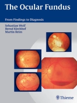
Learn how to ‘read’ the optic fundus: What tests are indicated? How do I interpret the findings? What is the next step? This book guides you quickly and confidently from finding to diagnosis. Practice-oriented Organized by presentation Systematic listing of diagnoses for each presentation Sidebars with a brief summary of the signs and symptoms for each diagnosis Quick reference and study guide in one Comprehensive Describes various examination method Covers even rare findings Differential diagnosis Figures to illustrate each diagnosis Notes on appropriate treatment Confidence Learn to take prompt, goal-directed action. Apply various diagnostic options appropriately and economically. Gain confidence in dealing with equivocal findings.
قائمة المحتويات
<p><strong>1 Introduction</strong><br><strong>2 Examination Methods</strong><br>History<br>Functional Tests<br>Ophthalmoscopy<br>Objective Imaging Studies and Their Evaluation<br>Electrophysiologic Studies<br>Ultrasound Studies<br>Biopsy<br><strong>3 Appearance of Retinal and Choroidal Disorders</strong><br>Retinopathies with Focal or Mottled Lesions<br>Prominence of the Macula<br>Proliferation, Scarring, and Holes in the Macula<br>Depigmented and Pigmented Focal Lesions in the Macula<br>Macular Dystrophies with Mottled Lesions<br>Large Areas of Yellowish-White Exudative Retinopathy<br>Scattered Pigment Changes with Large Areas of Retinal and Choroidal Dystrophy<br>Peripheral Retinal and Choroidal Lesions<br>Retinal Detachments<br>Retinal Tumors<br><strong>4 Appearance of Vascular Disorders</strong><br>Ophthalmoscopic Structure of Fundus Vessels<br>Variants in the Course of the Retinal Vessels<br>Abnormal Vessels in the Retina<br>Rarefied and Elongated Vessels<br>Narrowed Arterioles and Congested Veins<br>Bleeding, Cotton-Wool Spots, Hard Exudates, and Retinal Edema<br>Capillary Aneurysms, Hard Exudates, Bleeding, and Neovascularization<br>Disorders Involving Primarily Retinal Bleeding<br>Peripheral Neovascularization of the Retina<br>Perivascular Infiltrates, Vascular Obliteration, and Retinal Bleeding in Inflammatory Vascular Disorders<br><strong>5 Phenomenology in Diseases of Vitreous Body</strong><br>Symptoms with Vitreous Opacities and Specific Examinations<br>Leukocoria<br>Blood Vessels in the Vitreous Body<br>Small Opacities in the Vitreous Body<br>Diffuse Opacities in the Vitreous Body<br>Large Opacities in the Vitreous Body<br><strong>6 Appearance of Optic Nerve Disorders</strong><br>Blurred Appearance, Hyperemia, and Protrusion—Optic Disc Edema<br>Pale, Often White, Sharply Demarcated Optic Disc—Optic Nerve Atrophy<br>Excavations of the Optic Nerve<br>Anomalous Tissue On and Adjacent to the Optic Disc<br><strong>7 Diseases without Conspicuous Changes of the Fundus</strong><br>Floaters<br>Unilateral Visual Impairment in Children—Amblyopia<br>Acute Visual Impairment with Normal Optic Disc<br>Color Vision Defects<br>Incipient Tapetoretinal Degeneration<br>Congenital Stationary Night Blindness (CSNB)<br>Night Blindness with Hereditary Deficiency of the Retinol-Binding Protein<br>Stimulation<br>Visual Agnosia</p>
عن المؤلف
Sebastian Wolf, Bernd Kirchhof, Martin Reim


