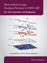
As its title suggests, this innovative book has been written for life scientists needing to analyse their data sets, and programmers, wanting a better understanding of the types of experimental images life scientists investigate on a regular basis. Each chapter presents one self-contained biomedical experiment to be analysed. Part I of the book presents its two basic ingredients: essential concepts of image analysis and Matlab. In Part II, algorithms and techniques are shown as series of ‘recipes’ or solved examples that show how specific techniques are applied to a biomedical experiments like Western Blots, Histology, Scratch Wound Assays and Fluoresence. Each recipe begins with simple techniques that gradually advance in complexity. Part III presents some advanced techniques for the generation of publication quality figures. The book does not assume any computational or mathematical expertise.
* A practical, clearly-written introduction to biomedical image analysis that provides the tools for life scientists and engineers to use when solving problems in their own laboratories.
* Presents the basic concepts of MATLAB software and uses it throughout to show how it can execute flexible and powerful image analysis programs tailored to the specific needs of the problem.
* Within the context of four biomedical cases, it shows algorithms and techniques as series of ‘recipes’, or solved examples that show how a particular technique is applied in a specific experiment.
* Companion website containing example datasets, MATLAB files and figures from the book.
Tabla de materias
Preface vii
Acknowledgements ix
About the Companion Website xi
1 The Basic Ingredients 1
1.1 The Matlab Environment 1
1.2 Introduction to Matlab 3
1.3 Operations with Matrices 7
1.4 Combining Matrices 10
1.5 Addressing a Matrix 13
1.6 Mathematical Functions and Graphical Display 17
1.7 Random Numbers 23
1.8 Statistics in Matlab 26
1.9 Displaying Two-Dimensional Matrices 29
1.10 Scripts Functions and Shortcuts 37
1.11 Using Help 43
2 Introduction to Images 45
2.1 An Image as a Matrix 45
2.2 Reading Images 46
2.3 Displaying Images 49
2.4 Colormap 54
2.5 Thresholding and Manipulating Values of Images 59
2.6 Converting Images into Doubles 68
2.7 Save Your Code and Data 69
3 Introduction to Colour 71
3.1 Mixing and Displaying Colours 71
4 Western Blots 79
4.1 Recipe 1: Many Ways to Display a Western Blot 80
4.2 Recipe 2: Investigating the Numbers That Make a Western Blot 93
4.3 Recipe 3: Image Histograms 97
4.4 Recipe 4: Transforming an Image of a Western Blot 104
4.5 Recipe 5: Quantification of the Data 111
4.6 Recipe 6: Investigating Position of Bands 121
5 Scratch Wound Assays 135
5.1 Analysis of Scratch Wound Assays 135
5.2 Recipe 1: Low Pass Filtering Scratch Wound Assays in the Spatial Domain 139
5.3 Recipe 2: High Pass Filtering Scratch Wound Assays in the Spatial Domain 143
5.4 Recipe 3: Combining Filters and Morphological Operations 154
5.5 Recipe 4: Sensitivy to Thresholds and Hysteresis Thresholding 161
5.6 Recipe 5: Morphological Operators 167
5.7 Recipe 6: Measuring Distances Between Cellular Boundaries 178
5.8 Recipe 7: Introduction to Fourier Analysis 187
5.9 Recipe 8: Filtering Scratch Wound Assays in the Fourier Domain 201
References 213
6 Bright Field Microscopy 215
6.1 Recipe 1: Changing the Brightness and Contrast of an Image 215
6.2 Recipe 2: Shading Correction: Estimation of Shading Component as a
Plane 224
6.3 Recipe 3: Estimation of Shading Component with Filters Morphological
Operators and Envelopes 235
6.4 Recipe 4: Mosaicking and Stitching 247
6.5 Recipe 5: Pixel Intensity and Histograms in Immunohistochemistry 261
6.6 Recipe 6: Hue-Saturation-Value 271
6.7 Recipe 7: Multidimensional Histograms 278
Reference 289
7 Fluorescence Microscopy 291
7.1 Recipe 1: Separating and Combining Colour Channels 294
7.2 Recipe 2: Investigating the Scaling of Values 298
7.3 Recipe 3: Automatic Threshold Selection 301
7.4 Recipe 4: Measuring Absolute and Relative Areas 304
7.5 Recipe 5: Counting Nuclei 305
7.6 Recipe 6: Quantification of Region Properties Beyond the Area 308
7.7 Recipe 7: Dividing an Image into Regions 310
7.8 Recipe 8: Batch Processing and Montages 316
7.9 Recipe 9: A Myriad of Measurements 327
References 341
8 Creating Publication-Quality Figures from Matlab 343
8.1 Recipe 1: Modifying the Characteristics of the Figures 344
8.2 Recipe 2: Numerous Plots in One Figure 352
8.3 Recipe 3: Three-Dimensional Ribbons with Different Annotations 362
8.4 Recipe 4: Three-Dimensional Graphics 378
8.5 Recipe 5: Projections 388
8.6 Recipe 6: Four-Dimensional Data Set Explored 391
Index 401
Sobre el autor
Constantino Carlos Reyes-Aldasoro (BSEE UNAM, Mexico, MSc Imperial College London, UK, Ph D Warwick, UK) is an interdisciplinary scientist with skills that span from microscopy to image analysis of biomedical data sets to advanced computer programming. His teaching experience is also wide ranging from Engineering, Physics and Computing to Medicine and Radiology. His research concentrates on Biomedical Image Analysis of Cancer, Microcirculation and Inflammation. He has published numerous journal/conference papers in both Biological and Engineering fields, which have been cited in more than 500 publications. He is a senior member of the Institute of Electrical and Electronics Engineers and executive committee member of the British
Association for Cancer Research.
