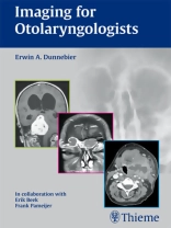
A practical imaging primer designed specifically for ENTs
Imaging for Otolaryngologists distils the essentials of otolaryngologic imaging into a concise reference that concentrates on key topics that are of immediate interest to otolaryngologists practicing in a modern clinical environment.
Prepared by a renowned otolaryngologist, and reviewed and supplemented by expert radiologists, the book provides a well-rounded perspective. The central focus is on image interpretation, including the disease-specific characteristics, the features necessary for successful diagnosis, and the implications for surgery. Each of the 465 high-quality images is clearly labeled, and where appropriate comparisons are made between CT scans and MR images to show complementary functions and limitations.
Imaging for Otolaryngologists helps readers:
- Evaluate the cross-sectional anatomy in rhinology, otology, and laryngology on plain films, CT scans, and MR images
- Appreciate the contribution and limitations of plain films, CT, and MRI in the management of otolaryngologic diseases
- Select the best imaging modality for chronic, acute, and emergency otolaryngologic conditions
- Understand which radiological appearances to look for in the diagnosis of common and less common otolaryngologic diseases
विषयसूची
General
1 Radiographic Imaging Techniques and Interpretation
Temporal Bone
2 Radiologic Anatomy of the Temporal Bone
3 Pathology of the Temporal Bone
Skull Base
4 Radiologic Anatomy of the Skull Base
5 Pathology of the Skull Base
Nose
6 Radiologic Anatomy of the Nasal Cavity and Paranasal Sinuses
7 Pathology of the Nasal Cavity and Paranasal Sinuses
Neck
8 Radiologic Anatomy of the Neck
9 Pathology of the Neck


