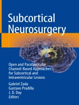
Our understanding of human neuro-anatomy, and ability to safely access lesions in complex locations, are in continuous evolution. The subcortical white matter space is among the most intricate, yet least understood, regions of the brain, with regards to its billions of connections and the subtle clinical and clinical functions it subserves. Neurosurgical procedures in the subcortical space and intraventricular system have been traditionally very difficult due to their depth, the need for brain retraction, and limited understanding and imaging capability of this region. Common lesions encountered in the subcortical space include brain metastases, gliomas, and intracerebral hemorrhage. Surgical access to this region has classically been hindered, and is highly limited by evolving technological applications to medicine and surgery. Traditionally, the technology (optics, imaging, resection devices, illumination) needed to perform safe subcortical surgery was not commensurate with the surgeon’s needs.
Over the past decade, major strides in our ability to image, navigate, and safely access subcortical tumors and other lesions have been made. These include parafascicular, trans-sulcal approaches that may be channel-based to provide safe retraction of the cortical and subcortical matter. A confluence of optical, computational and mechanical technology have greatly enabled our ability to treat such lesions, and include advanced MR imaging such as diffusion tractography, neuronavigation, channel-based access ports, exoscopic visualization, fiber-optic illumination, and novel resection devices.
Parafascicular, channel-based subcortical surgery is a relatively new field with evolving indications and approaches that promises to evolve considerably over the next several decades. We aim to develop the first comprehensive reference text compiling the known evidence and experience from expert practitioners in the field of subcortical parafascicular surgery. This book will provide a major foundation for future development of the field, and be a first and definitive reference for decades to come.
Subcortical Neurosurgery: Open and Parafascicular Channel-Based Approaches for Subcortical and Intraventricular Lesions will be the definitive reference on surgery of the subcortical region. It will comprehensively discuss all aspects of treatment of subcortical and intraventricular lesions, including neuroanatomy and neuroimaging of the subcortical space, principles of parafascicular subcortical channel-based surgery, common indications and approaches, and focused chapters for common subcortical lesions.
The first section of the reference focuses on the intricate anatomy and neuro-imaging of the subcortical space and ventricular system, with special emphasis on intricate white matter tracts and diffusion tractography imaging. The next section of the book discusses principles of both open and parafascicular, channel-based approaches to subcortical and intraventricular lesions, in addition to workhorse approaches to common subcortical compartments. Finally, specific pathological subcortical lesions that can be commonly addressed via parafascicular channel-based approaches, including brain metastases, gliomas, and intracerebral hemorrhage will be addressed. Authored by experts in the field of subcortical neurosurgery, this book was developed to provide a unique, comprehensive text for neurosurgeons, neuro-radiologists, and trainees from a variety of specialties interested in evolving minimally disruptive access and treatment of the subcortical space.
विषयसूची
The subcortical space: anatomy of the subcortical white matter.- Common pathology of the subcortical region.- The ventricular system: Anatomy and common lesions.- Advanced neuro-imaging of the subcortical space.- Advanced neuro-imaging of the human ventricular system.- The evolution of trans-sulcal, channel-based parafascicular surgery.- Open transcortical approaches to subcortical lesions.- Traditional open and neuro-endoscopic approaches to intraventricular pathology.- Trans-sulcal, channel-based parafascicular surgery: Technological considerations and instrumentation.- Standard parafascicular approaches to subcortical regions.- Trans-sulcal, channel-based parafascicular surgery: Basic concepts and a general overview.- Trans-sulcal, channel-based parafascicular surgery for brain metastases.- Trans-sulcal, channel-based parafascicular surgery for gliomas and other primary, intrinsic subcortical tumors.- Trans-sulcal, channel-based parafascicular biopsy techniques.- Trans-sulcal, channel-based parafascicular surgery for intraventricular lesions.- Trans-sulcal, channel-based parafascicular surgery for intracerebral hematoma (ICH).- Trans-sulcal, channel-based parafascicular surgery for cavernous angiomas and other vascular lesions.- Trans-sulcal, channel-based parafascicular surgery for tumors; Adjunctive measures and techniques.- Case selection and surmounting the learning curve.- Future directions.
लेखक के बारे में
Gabriel Zada, MD, MS
Professor of Neurosurgery, Otolaryngology, and Medicine
Co-Director, USC Pituitary Center
Co-Director, USC Radiosurgery Center
Neuro-Oncology and Endoscopic Pituitary/Skull Base Program
Keck School of Medicine of USC
1200 N. State Street #3300
Los Angeles, CA 90033
Office: (323) 442-6290
Gustavo Pradilla, M.D.
Assistant Professor
Department of Neurological Surgery
Emory University School of Medicine
Chief of Neurosurgery Service – Grady Memorial Hospital
Director Cerebrovascular Research Laboratory
1365 Clifton Rd, NE
Building B, Suite 2200
Atlanta, GA 30322
Phone: 404-778-5770 Fax: 404-778-3279


