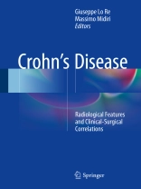
This book describes and illustrates the radiological findings that are characteristic of Crohn’s disease and correlates these appearances with clinical and surgical findings in order to assist the reader in implementing an appropriate diagnostic approach in this complex disease, which often differs from patient to patient. All of the modern imaging modalities and techniques are covered, including ultrasound, multidetector row computed tomography (MDCT), magnetic resonance imaging (MRI), and combinations of MDCT and MRI with enteroclysis and enterography. The current value of X-ray and the role of endoscopy are also examined. Care is taken to cover specific patient groups, including pediatric patients, those with perianal disease or anal fistulas, and patients who have undergone surgery. In addition to surgical approaches, including minimally invasive options, other new therapies are discussed. The book will be invaluable for diagnostic radiologists, gastroenterologists, and surgeons.
Daftar Isi
Epidemiologic aspect of Crohn’s Disease.-Clinical presentation of Crohn’s disease.- Laboratory tests in Crohn’s disease.- Decourse of Disease.-Diagnostic and therapeutic role of endoscopy in Crohn’s disease.- Perianal Crohn’s.- Standard therapeutic approach and new therapies.- Current status of imaging in small and large bowel diseases.- X-ray in inflammatory and bowel disease, Jurassic technique or yet still useful in the 21st century?.- US in Inflammatory Bowel Disease.- MDCT enteroclysis and enterography.- MRI enteroclysis and enterography.- Anal fistula in Crohn’s disease.- Extraintestinal findings in Crohn’ Disease patients.- Follow- up of IBD.- Surgery and Crohn’s disease.- Surgical emergencies in Crohn’s Disease.- Perianal surgery in in Crohn’ Disease.
Tentang Penulis
Massimo Midiri is Full Professor of Radiology and Director of the Section of Radiological Sciences, Department of Biopathology and Medical and Forensic Biotechnologies, University Hospital “Paolo Giaccone”, Palermo, Italy, which hosts a strong school of surgical oncology. In recent years, Dr. Midiri has succeeded in establishing a new lab for preclinical studies at the Section of Radiological Sciences, with installation of a very high magnetic field strength (7T) MRI scanner for small animals. The Section of Radiological Sciences also hosts the first and only Italian transcranial MRg FUS system for the treatment of neurological disorders. Dr. Midiri is lead author or co-author of more than 600 publications indexed on Google Scholar, more than 250 of which are indexed in Pub Med.
Giuseppe Lo Re is Adjunct Professor at the School of Specialization in Medical Imaging, Faculty of Medicine and Surgery, University of Palermo, Italy. After graduating in Medicine and Surgery in 2002 (110/110 and Thesis with Honors), Dr. Lo Re completed his specialization in Radiology in 2006 (50/50), and in 2011 gained his Ph D from the University of Palermo. His interests include study of intestinal diseases using conventional radiology, ultrasound, CT, and MRI and study of perianal disease using MRI. He is lead or co-author of 200 scientific publications, including 13 book chapters and 38 scientific articles in national and international journals.


