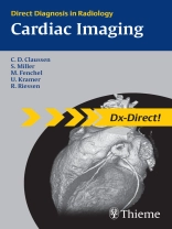
Dx-Direct is a series of eleven Thieme books covering the main subspecialties in radiology. It includes all the cases you are most likely to see in your typical working day as a radiologist. For each condition or disease you will find the information you need — with just the right level of detail.
Whether you are a resident or a trainee, preparing for board examinations or just looking for a superbly organized reference, Dx-Direct is the high-yield choice for you!
The series covers the full spectrum of radiology subspecialties including:
- Brain
- Gastrointestinal
- Cardiac
- Breast
- Urogenital
- Vascular
- Spinal
- Head and Neck
- Musculoskeletal
- Pediatric
- Thoracic
Dx-Direct gets to the point: Definitions, Epidemiology, Etiology, and Imaging Signs Typical Presentation, Treatment Options, Course and Prognosis Differential Diagnosis, Tips and Pitfalls, and key References, all combined with high-quality diagnostic images.
Jadual kandungan
1 Ischemic Heart Disease
Coronary Heart Disease (CHD)
Unstable Angina Pectoris
Acute Myocardial Infarction (AMI)
Chronic Myocardial Infarction
Ventricular Aneurysm
Syndrome X
Prinzmetal Angina
Aortocoronary Bypass Surgery
Coronary Anomalies
Bland–White–Garland Syndrome
Kawasaki Syndrome
2 Heart Failure
Acute Heart Failure
Chronic Heart Failure
Heart Transplantation
3 Acquired Valvular Heart Diseases
Aortic Stenosis
Aortic Insufficiency
Mitral Stenosis
Mitral Insufficiency
Pulmonary Stenosis
Pulmonary Insufficiency
Tricuspid Stenosis
Tricuspid Insufficiency
Combined Valvular Disease
Mitral Valve Prolapse
Prosthetic Heart Valves
Ross Aortic Valve Replacement
Aortic Valve Reconstruction
4 Cardiomyopathy
Dilated Cardiomyopathy (DCM)
Hypertrophic Cardiomyopathy (HCM)
Arrhythmogenic Right Ventricular Cardiomyopathy
Restrictive Cardiomyopathy (RCM)
Unclassified Cardiomyopathies (ILNC)
Apical Ballooning
Sarcoidosis
Amyloidosis
Hemochromatosis, Hemosiderosis
Uremic Cardiomyopathy
Toxic Cardiomyopathy
5 Inflammatory Heart Diseases
Myocarditis
Acute Pericarditis
Constrictive Pericarditis
Infectious Endocarditis
Hypereosinophilic Syndrome (Löffler Endocarditis)
Postinfarction Pericarditis, Dressler Syndrome
6 Hypertension
Arterial Hypertension
Chronic Pulmonary Hypertension
Acute Pulmonary Hypertension, Pulmonary Embolism
7 Tumors and Other Masses
Thrombus
Myxoma
Lipoma
Fibroma
Papillary Fibroelastoma
Pericardial Cyst
Metastases
Angiosarcoma
Undifferentiated Sarcoma
Rhabdomyosarcoma
Lymphoma
Pulmonary Artery Sarcoma
Mediastinal Tumors
8 Trauma
Cardiac Contusion
Aortic Rupture
Coronary Dissection
Pulmonary Artery Rupture
9 Congenital Heart Diseases
Atrial Septal Defect (ASD)
Ventricular Septal Defect (VSD)
Eisenmenger Syndrome
Patent Ductus Arteriosus
Bicuspid Aortic Valve
Coarctation of the Aorta
Aortic Arch Anomalies
Heterotaxy Syndrome
Tricuspid Atresia
Truncus Arteriosus
Cor Triatriatum
Scimitar Syndrome
Atrioventricular Septal Defect (AVSD)
Ebstein Anomaly
Tetralogy and Pentalogy of Fallot
Hypoplastic Left Heart Syndrome (HLHS, Single Ventricle)
Pulmonary Atresia
Transposition of the Great Arteries (dextro-TGA)
Corrected Transposition of the Great Arteries (levo-TGA)
Double-Outlet Right Ventricle (DORV)
Total Anomalous Pulmonary Venous Connection (TAPVC)
Fontan Operation
Blalock-Taussig Shunt
10 Diseases of the Great Vessels
Aortic Aneurysm
Aortic Ectasia
Aortic Dissection
Carotid Stenosis
Subclavian Steal Syndrome
Thoracic Outlet Syndrome (TOS)
Takayasu Arteritis
11 Standard Views of the Heart
Where are Structures Located in the Chest Radiograph?
Standard Views of the Heart: Overview
Two Chamber Long-Axis View Parallel to the Septum, LV
Short-Axis View
Four-Chamber View
LVOT
LVOT, ‘Three-Chamber View’
Two-Chamber Long-Axis View Parallel to the Septum, RV
RVOT
12 Appendix
Normal Values (Adults)
Classifications of Myocardial Segments
Segmental Divisions of the Coronary Arteries
Classification of Pulmonary Venous Drainage Patterns
NYHA Criteria for Symptoms of Heart Failure
Classification of Thoraco-abdominal Aortic Aneurysms


