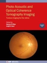
This book covers the state-of-the-art techniques of fundus imaging for the diagnosis of retinal diseases. It is part of a three-volume work that describes the latest imaging techniques in which to bring optical coherence tomography (OCT), fundus Imaging and optical coherence tomography angiography (OCTA) to accurately facilitate the diagnosis of retinal diseases. Clinical disorders of the retina have been attracting the attention of researchers, aiming at reducing the blindness rate. This includes uveitis, diabetic retinopathy, macular edema, endophthalmitis, proliferative retinopathy, age-related macular degeneration and glaucoma. Treatment is significantly dependent on having early and accurate diagnosis, which can be significantly improved by employing the techniques described in the book.
Key Features
- Provides a comprehensive overview of all pertinent topics related to fundus imaging techniques, applicable to diagnosis of eye disorders
- Offers a unique coverage of Neural Networks in distinguishing eye diseases
- Machine learning techniques are presented in detail throughout
- Many of the chapter contributors are world-class researchers
- Extensive references will be provided at the end of each chapter to enhance further study
Inhoudsopgave
1. Texture Interpretability of Fundus Imaging and Diabetic Retinopathy: A Review
2. A two-phase novel optic disc detection algorithm based on vesselness distribution and a fuzzy classifier using vessel ramification and fundus color features
3. Application of enface image registration / alignment to introduce new ocular imaging biomarkers
4. Existing techniques used for retinal image analysis for automated detection and prediction of AMD
5. Interobserver variability in the determination of diabetic retinopathy and quality of fundus image
6. Computer aided diagnosis of plus disease in preterm infants
7. Retinal disease management using fundus autofluorescence images
8. A review of mainstream ophthalmic AI algorithms: Advances, limitations and challenges
9. Diabetic Retinopathy detection and classification through fundus images using Alexnet
10. An Approach for Disclosing of the Optic Disk and Detection of Exudates in fundus images
11. A framework for joint Cup and Disc Segmentation in Fundus Images
Over de auteur
Ayman El-Baz is a Distinguished Professor at University of Louisville, Kentucky, United States and University of Louisville at Alamein International University (Uof L-AIU), New Alamein City, Egypt. Dr. El-Baz has almost two decades of hands-on experience in the fields of bio-imaging modeling and non-invasive computer-assisted diagnosis systems. He has authored or co-authored more than 700 technical articles (213 journal articles, 53 books, 107 book chapters, 262 refereed-conference papers, 216 abstracts, and 38 US patents and Disclosures).
Jasjit S. Suri is an innovator, scientist, and an internationally known world leader in biomedical engineering. Dr. Suri has spent over 25 years in the field of biomedical engineering/devices and its management. Dr. Suri was crowned with President’s Gold medal in 1980 and made Fellow of the American Institute of Medical and Biological Engineering for his outstanding contributions. In 2018, he was awarded the Marquis Life Time Achievement Award for his outstanding contributions and dedication to medical imaging and its management.


