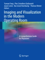
This text provides a state of the art overview of tools for guiding surgeons in the modern operating room. The text explains how many modalities in the current armamentarium of radiologic imaging have been brought to the operating room for real time use. It also explains the current use of near infrared, fluorescent, and chemo-luminescent imaging to guide minimally invasive and open surgery to improve outcome. The book is separated into two sections. The first, discusses the biologic principles that underlie novel visualization of normal organs and pathology. The currently available equipment and equipment anticipated in the near future is covered. The second section summarizes current clinical applications of advanced imaging and visualization in the OR. Novel means of visualizing normal anatomic structures such as nerves, bile duct, and vessels that enhance safety of many operations are covered. Novel biologic imaging using radio-labeled and fluorescent-labeled molecular probes that allow identification of inflammation, vascular abnormalities, and cancer are also discussed.
Authored by scientists who pioneer research in optics and radiology, tool makers who use this knowledge to make surgical equipment, and surgeons who innovate the field of surgery using these new operative tools, Imaging and Visualization in the Modern Operating Room is a valuable guide for surgeons, residents and fellows entering the field.
Inhoudsopgave
Basis of practice.- Lighting in the Operative Room: Current Technologies and Considerations.- Modalities for Intraoperative Imaging.- Fluorescents Probes.- Detectors for Intraoperative Molecular Imaging: From Probes to Scanners.- Isotopes and Procedural Imaging.- Radiologically Imageable Nanoparticles.- Flat Panel CT and the Future of OR imaging and Navigation.- Cerenkov Luminescence Imaging.- Organ Deformation and Navigation.- Clinical Milestones for Optical Imaging.- 3D in the Minimally Invasive Surgery (MIS) Operating Room: Cameras and Displays in the Evolution of MIS.- Molecular Tumor Marker Recognition: Nanotechnology for On-Line Diagnostics in the OR.- Ultra Small Fluorescent Silica Nanoparticles as Intraoperative Imaging Tools for Cancer Diagnosis and Treatment.- Image Processing Technologies for Motion Compensation.- Current Clinical Applications.- Near Infra-red Imaging in Robotic Surgery.- New Preoperative Images, Surgical Planning and Navigation.- New Generation Radiosurgery.- Breast Lesion Localization.- Intraoperative Breast Imaging and Image-Guided Treatment Modalities.- Sentinel Lymph Node Mapping: Current Practice and Future Developments.- Narrow Band Cystoscopy.- Fluorescence Imaging of Human Bile and Biliary Anatomy.- PET Guided Interventions from Diagnosis to Treatment.
Over de auteur
Yuman Fong, M.D.
Department of Surgery
City of Hope National Medical Center
Duarte, CA, USA
Pier Cristoforo Giulianotti, M.D.
Department of Surgery
University of Illinois Hospital and Health Sciences System
Chicago, IL, USA
Jason S. Lewis, Ph.D.
Department of Radiology
Memorial Sloan-Kettering Cancer Center
New York, NY, USA
Bas Groot Koerkamp, M.D.
Department of Surgery
Erasmus University Medical Center
Rotterdam, The Netherlands
Thomas Reiner, Ph.D.
Department of Radiology
Memorial Sloan Kettering Cancer Center
New York, NY, USA


