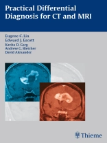
A concise reference for everyday practice
Practical Differential Diagnosis in CT and MRI is a one-stop resource for the differential diagnosis of common and rare radiologic findings and conditions in all regions of the body. For each finding and diagnosis, the book provides a complete list of differential diagnoses as well as the features that will help the clinician differentiate diseases with similar findings.
Highlights:
- Concise descriptions aid the identification of key radiologic signs
- Easy-to-use tables and bullet-point lists facilitate rapid review of important information about findings, differentiating features, and disease entities
This pocket-sized book is ideal for residents preparing for board examinations as well as for radiologists in practice.
Tabela de Conteúdo
BRAIN
Tumor and Tumor-like Lesions
1. Peripherally Enhancing Cystic Brain Lesions
2. Metastases versus Primary Brain Neoplasms
3. Intraventricular Neoplasms
4. Tumefactive Demyelination
5. Posterior Fossa Neoplasms in Children
6. Posterior Fossa Cystic Neoplasms
7. Cerebellopontine Angle Lesions
8. Posterior Fossa Cysts and Cerebellar Malformations
9. Meningeal Enhancement
10. Sellar and Parasellar Lesions
11. Dermoid Cysts and Lipomas
12. Pineal Region Lesions
Vascular
13. Arterial Infarcts versus Venous Infarcts
14. Bilateral Thalamic Lesions
15. Intracranial Hemorrhages
16. Perivascular Spaces versus Lacunar Infarcts
Infection
17. Intracranial Manifestations of Human Immunodeficiency Virus
Miscellaneous
18. Hyperdense and Calcified Lesions on Computed Tomography
19. Corpus Callosum Lesions
20. Abnormal Basal Ganglia Signal on Magnetic Resonance Imaging
21. Hydrocephalus
22. Restricted Diffusion
HEAD AND NECK
Skull
23. Focal Calvarial Lesions
24. Diffuse Calvarial Lesions
Skull Base
25. Skull Base Lesions
26. Jugular Foramen Lesions
Sinonasal Region
27. Sinonasal Lesions
28. Abnormal Sinus Density and Signal
Orbit
29. Orbital Vascular Lesions
30. Orbital Lymphoproliferative and Inflammatory Disorders
31. Optic Nerve-Sheath Complex Lesions
32. Lacrimal Gland Lesions
Salivary Gland
33. Salivary Gland Lesions
Thyroid Lesions and Calcifications
34. Thyroid Diseases and Lesions
Carotid Space
35. Carotid Space Masses
Congenital Cystic Neck Masses
36. Congenital Cystic Neck Masses
Lymph Nodes
37. Lymph Node Disease
Ear and Temporal Bone
38. Ear and Temporal Bone Lesions
Larynx
39. Vocal Cord Lesions and Paralysis
Carcinomas and Neoplasms
40. Head and Neck Cancer
41. Lymphoma
42. Radiation Changes
CHEST
Patterns and Signs
43. The Mosaic Pattern of Lung Attenuation
44. The Tree-in-Bud Pattern
45. Ground-Glass Opacity
46. The Halo Sign
47. The Crazy-Paving Pattern
48. The Angiogram Sign
49. The Feeding Vessel Sign
Airways
50. Bronchiectasis
51. Tracheobronchomalacia
52. Asthma and Associated Conditions
53. Emphysema
Mediastinum
54. Differential Diagnosis of Mediastinal Masses Based on Common Sites of Origin
Pulmonary Parenchyma
55. High-Resolution Computed Tomography of Chronic
Diffuse Infiltrative Lung Disease
56. Idiopathic Interstitial Pneumonia
57. Cystic Lung Disease
58. Pneumoconiosis
59. Radiation and Drug-Induced Lung Disease
60. The Solitary Pulmonary Nodule
61. Lung Transplantation
62. Pleural Effusions
63. Pericardial Effusion
64. Pulmonary Embolism
65. Aortic Dissection
66. Cardiac Aneurysms and Abnormalities
ABDOMEN
Liver
67. Overview of Liver Lesions
68. Hypervascular Liver Lesions
69. Liver Lesions with Delayed/Prolonged Enhancement
70. Liver Lesions with Central Scar
71. Cystic Liver Lesions
72. Calcified Liver Lesions
73. Hemorrhagic Liver Lesions
74. Liver Lesions with Macroscopic Fat
75. Liver Lesions with Microscopic Fat
76. Liver Lesions with Hyperintensity on T1-Weighted
Magnetic Resonance Imaging
77. Liver Lesions with Hypointensity on T2-Weighted
Magnetic Resonance Imaging
78. Transient Hepatic Attenuation Difference
79. Hepatic Capsular Retraction
80. Periportal Halo
81. Liver Lesions with Supraparamagnetic Iron Oxide Uptake
82. Diffuse Liver Disease
83. Cirrhosis
84. Mimics of Cirrhosis
Bile ducts
85. Magnetic Resonance Cholangiopancreatography Pitfalls
Pancreas
86. Cystic Pancreatic Lesions
87. Hypervascular Pancreatic Lesions
88. Focal Chronic Pancreatitis versus Pancreatic Cancer
89. Pancreatic Lymphoma versus Carcinoma
Spleen
90. Splenic Lesions
Adrenals
91. Adrenal Adenoma versus Metastasis
92. Adrenal Lesions with Macroscopic Fat
93. Cushing Syndrome
94. Hyperaldosteronism
Kidneys
95. Hyperdense Renal Lesions
96. Renal Lesions with Fat
97. Angiomyolipomas with Minimal Fat
98. Renal Parenchyma with Hypointensity on T2-Weighted Magnetic Resonance Imaging
99. Renal Infarction versus Pyelonephritis
Bowel
100. Imaging Signs of the Bowel
101. Diverticulitis versus Colon Cancer
102. Perforated Appendicitis
103. Epiploic Appendagitis versus Omental Infarction
Peritoneum/Retroperitoneum
104. Solid Peritoneal Masses
105. Cystic Peritoneal and Retroperitoneal Masses
106. Peritoneal Calcification
107. Retroperitoneal Fibrosis
PELVIS
Uterus
108. The Differential Diagnosis of Leiomyomas
109. Uterine Sarcomas
110. Endometrial Carcinoma versus Polyp
111. Septate versus Bicornuate Uterus
Ovaries
113. Benign versus Malignant Ovarian Mass
114. Ovarian Lesions with Hyperintensity on T1-Weighted Magnetic Resonance Imaging
115. Ovarian Lesions with Hypointensity on T2-Weighted Magnetic Resonance Imaging
Perineum
116. Periurethral and Vaginal Cysts
Prostate
117. Prostatic Cysts
MUSCULOSKELETAL
Bone lesions
118. Bone Lesions with Low Hypointensity on T2-Weighted Magnetic Resonance Imaging
119. Benign Bone Lesions with Surrounding Edema
120. Bone Lesions with Fluid-Fluid Levels
121. Enchondroma versus Chondrosarcoma
122. Osteochondroma versus Chondrosarcoma
Soft tissue lesions
123. Malignant versus Benign Soft Tissue Lesions
124. Cystic-Appearing Soft Tissue Masses
125. Soft Tissue Lesions with Fluid-Fluid Levels
126. Lipoma versus Liposarcoma
127. Nerve Sheath Tumors
Bone marrow
128. Osteomyelitis versus Neuropathic Arthropathy in the Diabetic Foot
129. Subchondral Bone Marrow Edema
Joints
130. Synovial Chondromatosis versus Rice Bodies
131. Hypointense Synovium on T2-Weighted Magnetic Resonance Imaging
Knee
132. Increased Meniscal Signal Not Related to Tear
133. Meniscal Tear without Signal Extending to the Articular Surface
134. Absent Bow-Tie Sign
135. Meniscal Extrusion
136. Medial Fluid Collections around the Knee
137. Fluid Collections around the Posterior Cruciate Ligament
Shoulder
138. Increased Signal in the Rotator Cuff Not Related to Full-Thickness Tear
139. Superior Labral Anterior-Posterior Tear versus Sublabral Recess
140. Abnormal Signal in the Rotator Cuff Muscles
SPINE
Vertebrae
141. Focal Vertebral Lesions
142. Diffusely Abnormal Marrow Signal on Magnetic Resonance Imaging
143. Benign versus Pathologic (Neoplastic) Fracture
144. Posterior Element Lesions
145. Posterior Vertebral Body Scalloping
Spinal Canal Lesions
146. Lesions within the Spinal Canal
147. Intradural Extramedullary Lesions
148. Intramedullary Spinal Cord Lesions
Infectious and Degenerative Processes
149. Diskitis/Osteomyelitis, Degenerative Disease, and Scar


