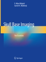
This book is a comprehensive guide to skull base imaging. Skull base is often a “no man’s land” that requires treatment using a team approach between neurosurgeons, head and neck surgeons, vascular interventionalists, radiotherapists, chemotherapists, and other professionals. Imaging of the skull base can be challenging because of its intricate anatomy and the broad breadth of presenting pathology. Although considerably complex, the anatomy is comparatively constant, while presenting pathologic entities may be encountered at myriad stages. Many of the pathologic processes that involve the skull base are rare, causing the average clinician to require help with their diagnosis and treatment. But, before any treatment can begin, these patients must come to imaging and receive the best test to establish the correct diagnosis and make important decisions regarding management and treatment. This book provides a guide to neuoradiologists performing that imaging and as a reference for relatedphysicians and surgeons.
The book is divided into nine sections: Pituitary Region, Cerebellopontine Angle, Anterior Cranial Fossa, Middle Cranial Fossa, Craniovertebral Junction, Posterior Cranial Fossa, Inflammatory, Sarcomas, and Anatomy. Within each section, either common findings in those skull areas or different types of sarcomas or inflammatory conditions and their imaging are detailed. The anatomy section gives examples of normal anatomy from which to compare findings against. All current imaging techniques are covered, including: CT, MRI, US, angiography, CT cisternography, nuclear medicine and plain film radiography. Each chapter additionally includes key points, classic clues, incidence, differential diagnosis, recommended treatment, and prognosis.
Skull Base Imaging provides a clear and concise reference for all physicians who encounter patients with these complex and relatively rare maladies.
Cuprins
Section I: Pituitary Region.- Rathke’s Cleft Cyst.- Pituitary Adenoma.- Craniopharyngioma.- Ectopic Neurohypohysis.- Hypothalamic Hamartoma.- Neurohypophyseal Sarcoidosis.- Intrasellar Arachnoid Cyst.- Langerhans Cell Histiocytosis.- Section II: Cerebellopontine Angle.- Epidermoid.- CPA Meningioma.- Vestibular Schwannoma.- CPA Arachnoid Cyst.- Section III: Anterior Cranial Fossa.- Anterior Fossa Meningioma.- Inverted Papilloma.- Juvenile Nasal Angiofibroma.- Neuroendocrine Carcinoma.- Nasofrontal Encephalocele.- Sinonasal Melanoma.- Nasal Dermoid.- Nasal Glioma.- Paranasal Sinus Mucocele.- Paranasal Sinus Cancer.- Osteoma.- Giant Cell Tumor.- Fibrous Dysplasia.- SNUC.- CSF Leaks.- Esthesioneuroblastoma. Section IV: Middle Cranial Fossa.- Jugular Foramen Paraganglioma.- Clivus Chordoma.- Lymphatic Malformations.- Allergic Fungal Sinusitis.- Basal Encephalocele.- Cavernous Sinus Aneurysm.- Cholesterol Granuloma Petrous Apex.- Skull Base Lymphoma.- Adenoid Cystic Carcinoma.- Aneurysmal Bone Cyst.- Trigeminal Schwannoma.- Section V: Craniovertebral Junction.- AOJ Dislocation.- Paget’s Disease.- Section VI: Posterior Cranial Fossa [PCF].- Posterior Fossa Arachnoid Cyst.- Posterior Fossa Meningioma.- Section VII: Inflammatory.- Skull Base Osteomyelitis.- Invasive Fungal Sinusitis.- Section VIII: Sarcomas.- Chondrosarcoma.- Ewings Sarcoma.- Fibrosarcoma.- Rhabdomyosarcoma.- Osteosarcoma.- Section IX: Anatomy.- Skull Base and Facial Foramina
Despre autor
F. Allan Midyett, M.D.’s primary passion is teaching neuroradiology, and he has been involved in teaching residents and fellows for the last two decades at large teaching hospitals including Howard University Hospital in DC. He is a senior member of the ASNR and holds a current Certificate of Added Qualification in Neuroradiology by the American Board of Radiology. Dr. Midyett has authored more than 100 scientific publications including book chapters and peer reviewed articles. His first book Orbital Imaging, co-authored by Dr. Mukherji, has been well received and is currently published in English, Spanish and Portuguese.
Suresh K. Mukherji, M.D. is Clinical Professor at Marian University, Director of Head & Neck Radiology at Pro Scan Imaging, and Regional Medical Director at Envision Physician Services, Carmel, Indiana, USA. He is a recognized authority in Head & Neck and Neuroradiology and has been an active contributor to the neuroradiology literature byauthoring over 400 scientific manuscripts and book chapters and has written or edited 13 textbooks. He has been an invited speaker on nearly 400 occasions. His primary interests have been focused on investigating emerging metabolic and physiologic imaging techniques to evaluate head and neck cancer and to differentiate recurrent tumor from post-therapeutic changes in previously treated patients. These technologies include imaging with fluorodeoxyglucose analogues imaged with prototype Single Photon Emission Computed Tomography, Gamma Cameras, standard PET, and CT-PET. Other metabolic and physiologic imaging techniques he has investigated include Thallium-201, magnetic resonance spectroscopy, CT perfusion and CT spectral imaging.
