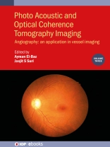
This book covers the state-of-the-art techniques of optical coherence tomography angiography (OCTA) imaging for the diagnosis of retinal diseases.
It is part of a three-volume work that describes the latest imaging techniques in which to bring optical coherence tomography (OCT), Fundus Imaging and optical coherence tomography angiography (OCTA) to accurately facilitate the diagnosis of retinal diseases. Clinical disorders of the retina have been attracting the attention of researchers, aiming at reducing the blindness rate. This includes uveitis, diabetic retinopathy, macular edema, endophthalmitis, proliferative retinopathy, age-related macular degeneration and glaucoma. Treatment is significantly dependent on having early and accurate diagnosis, which can be significantly improved by employing the techniques described in the book.
Key Features:
- Provides a comprehensive overview of all pertinent topics related to optical coherence tomography angiography (OCTA) imaging techniques, applicable to diagnosis of eye disorders.
- Offers a unique coverage of Neural Networks in distinguishing eye diseases.
- Machine learning techniques are presented in detail throughout.
- Many of the chapter contributors are world-class researchers.
- Extensive references will be provided at the end of each chapter to enhance further study.
Содержание
1. Clinical application of optical coherence tomography angiography in retinal diseases
2. Longitudinal optical coherence tomography and angiography of hyaloid vascular regression in developing mouse eyes
3. Stripe noise removal and vessel segmentation of octa images
4. Optical coherence tomography angiography changes in early type 3 neovascularization after anti-vascular endothelial growth factor treatment
5. Optical coherence tomography angiography in multiple sclerosis and neuromyelitis optica
6. Optical coherence tomography angiography for the diagnosis of polypoidal choroidal vasculopathy
7. Quantitative features for objective assessment of oct angiography
8. Clinical utility of OCT-Angio in Age-related Macular Degeneration
9. Optical Coherence Tomographic Angiography Imaging in Age-Related Macular Degeneration
10. Optical coherence tomography angiography in ophthalmology: an application in vessel imaging
11. Optical coherence tomography angiography: principles and clinical application
12. A Comprehensive Survey on Computer-Aided Diagnostic Systems in Diabetic Retinopathy Screening
Об авторе
Ayman El-Baz
Ayman El-Baz is a Distinguished Professor at University of Louisville, Kentucky, United States and University of Louisville at Al Alamein International University (Uof L-AIU), New Alamein City, Egypt. Dr. El-Baz earned his B.Sc. and M.Sc. degrees in electrical engineering in 1997 and 2001, respectively. He earned his Ph.D. in electrical engineering from the University of Louisville in 2006. Dr. El-Baz was named as a Fellow for Coulter, AIMBE and NAI for his contributions to the field of biomedical translational research. Dr. El-Baz has almost two decades of hands-on experience in the fields of bio-imaging modeling and non-invasive computer-assisted diagnosis systems. He has authored or co-authored more than 700 technical articles.
Jasjit S. Suri
Jasjit S. Suri, Ph D, MBA is a Fellow of IEEE, AIMBE, SVM, AIUM, and APVS. He is currently the Chairman of Athero Point, Roseville, CA, USA, dedicated to imaging technologies for cardiovascular and stroke. He has nearly ~22, 000 citations, co-authored 50 books, and has an H-index of 72.


