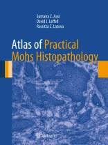
Mohs surgery is microscopically controlled surgery used to treat common types of skin cancer and allows for the removal of a skin cancer with a very narrow surgical margin and a high cure rate. However, for those involved with the Mohs procedure, it is critical to understand the optimal preparation and interpretation of frozen sections.
Complete with hundreds of high resolution figures, Atlas of Practical Mohs Histopathology is written by leading experts in the field and discusses everything from normal skin histology and rare tumors to pitfalls and incidental findings. Dermatologic surgeons, Mohs cutaneous surgeons, dermatopathologists and pathologists alike will find this book to be a comprehensive and indispensable reference.
สารบัญ
1. How to use this atlas.- 2. Normal skin histology.- 3. Basal cell carcinoma.- 4. Infiltrative basal cell carcinoma.- 5. Differentiating basal cell carcinoma from normal structures.- 6. Adnexal tumors.- 7. Microcystic adnexal carcinoma.- 8. Differentiating infiltrative basal cell carcinoma from other tumors.- 9. Squamous cell carcinoma in situ (Bowen’s disease) and actinic keratoses.- 10. Squamous cell carcinoma.- 11. Differentiating features of squamous cell carcinoma.- 12. Dermatofibrosarcoma protuberans.- 13. Rare tumors including atypical fibroxanthoma.- 14. Pitfalls and incidental findings.- 15. Artifacts.- 16. Bibliography.
เกี่ยวกับผู้แต่ง
David J. Leffell, MD David Paige Smith Professor of Dermatology, Otolaryngology, and Plastic Surgery Yale School of Medicine New Haven, Connecticut
Sumaira Z. Aasi, MD Associate Professor of Dermatology Stanford University School of Medicine Palo Alto, California
Rossitza Z. Lazova, MD Associate Professor of Dermatology and Pathology Yale School of Medicine New Haven, Connecticut


