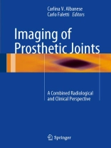
An increasing percentage of the population has at least one prosthetic joint. Imaging is required for both initial assessment and routine follow-up of the implant, and this book is intended as an informative and up-to-date guide to the subject. After an introductory section covering a range of background topics, the level of information offered by different imaging techniques in presurgical planning and postimplantation assessment is analyzed. The application of imaging to different joints is then carefully explored in chapters devoted to the foot and ankle, hip, knee, shoulder, and elbow, wrist and hand. In addition, two innovative chapters focus on periprosthetic DXA as the gold standard in monitoring implant survival and on the role of drug therapy in helping to ensure the durability of the prosthesis. A central feature of the book is its combination of the clinical and radiological perspectives; it will be of value to radiologists and orthopedic specialists, as well as residents inthese disciplines.
Mục lục
SECTION I Cellular elements, bone matrix, bone remodeling, osseointegration, implant material.- 1 Bone cells.- 2 Bone matrix proteins and mineralization process.- 3 Bone remodeling.- 4 Biomechanics and prosthetic osseointegration.- 5 Implant material.- SECTION II Images techniques, conventional RX, TC, RM, bone scintigraphy, periprosthetic DXA.- 6 Role of conventional RX, CT and MRI in the evaluation of prosthetic joints.- 7 Role of Nuclear Medicine in prosthesis surveillance.- 8 Periprosthetic DXA.- SECTION III Prosthetic joints of foot and ankle, hip, knee, shoulder, elbow, wrist and hand.- 9 Foot and ankle.- 10 Hip.- 11 Knee.- 12 Shoulder arthroplasty.- 13 Prosthetic joints of elbow, wrist and hand.- SECTION IV Drug therapy, bisphosphonate, teriparatide and parathormone, strontium ranelate.- 14 Drug therapy after implant.- 15 TBD- post surgery rehabilitation.


