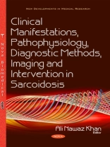
Sarcoidosis is a multisystem granulomatous disease of unknown etiology that is characterized by noncaseous epithelioid cell granulomas, which may affect almost any organ in the body. Intrathoracic involvement is common and accounts for most of the morbidity and mortality associated with this disease. The diagnosis is based on the total exclusion of other granulomatous disorders. The organs that are commonly involved are the lymph nodes, lungs, liver, spleen, skin, and eyes; these organs can be involved individually or in combination. The correlation of the clinical, radiological features along with the pathologic finding of non-caseating epithelioid cell granulomas is vital to establish the diagnosis. There is no single precise cause attributed to the causation of this disease. Genetic factors are suspected, due to the observation that racial groups such as African Americans, West Indians and Asians have a higher prevalence of sarcoidosis.Familial sarcoidosis is well-known, which may be attributed to genetic factors or the sharing of a similar environment. Environmental factors may also play a role by involving the uptake and processing of unknown antigens by the respiratory system. Occasional patients with sarcoidosis have an association with primary biliary cirrhosis, where the granulomas in both diseases look similar. Patients receiving treatment with anti-retroviral therapy or interferon alpha might have pulmonary granulomas as in HIV-infected patients and leukemia patients retrospectively. Sarcoidosis is more prevalent and is a more severe disease in blacks in the United States of America. Two-thirds of patients with sarcoidosis resolve spontaneously without specific treatment. Therapeutic measures, when required, rely on immune suppression. As the symptoms are varied in sarcoidosis, the differential diagnosis includes most non-specific systemic disorders. A chest radiograph (CXR) is usually the first diagnostic imaging study in patients with respiratory symptoms. A CXR is a non-invasive modality, widely available, easy to interpret and when correlated with the clinical findings, may be the only imaging required to diagnose pulmonary sarcoidosis. A CXR is also the most commonly used imaging technique for follow-up in patients with established diagnosis, and is reproducible and cost efficient. Conventional chest radiography, however, has its limitations. While it may be normal in 5-10% of patients with established sarcoidosis, in 25-30% of patients, the radiologic changes are nonspecific or atypical reducing the plain film sensitivity. In such cases, High-Resolution CT (HRCT) is useful in clarifying the diagnosis providing crucial information on the extent of the disease. Furthermore, HRCT, unlike a plain radiograph, can readily differentiate active inflammation from irreversible fibrosis.
Ali Nawaz Khan
Clinical Manifestations, Pathophysiology, Diagnostic Methods, Imaging and Intervention in Sarcoidosis [PDF ebook]
Clinical Manifestations, Pathophysiology, Diagnostic Methods, Imaging and Intervention in Sarcoidosis [PDF ebook]
购买此电子书可免费获赠一本!
格式 PDF ● 网页 219 ● ISBN 9781634858441 ● 编辑 Ali Nawaz Khan ● 出版者 Nova Science Publishers ● 发布时间 2016 ● 下载 3 时 ● 货币 EUR ● ID 7227083 ● 复制保护 Adobe DRM
需要具备DRM功能的电子书阅读器
